
Watch this 78-second video to see exactly what a sestamibi scan looks like—and why it only takes 25 minutes at our center, not hours like at other hospitals. No enclosed tubes, no stress, just fast and accurate imaging for parathyroid disease.
 Sestamibi scanning is the preferred way to localize diseased parathyroid glands prior to an operation. HOWEVER, sestamibi scans are wrong at least 50% of the time, even at the best places in the world. Be careful folks!
This scan was invented in the early 1990's and now is widely available at essentially every hospital in the United States. Sestamibi is a small protein which is labeled with the radio-pharmaceutical technetium-99. This very mild and safe radioactive agent is injected into the veins of a patient with parathyroid disease (hyperparathyroidism) and is absorbed by the overactive parathyroid gland. This is a very important concept--the parathyroid tumor will collect the radioactive dye. Furthermore, since normal parathyroid
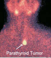
![]() glands are inactive when there is high calcium in the blood stream, they do NOT take up the radioactive particles. Therefore, a sestamibi scan will show the one bad parathyroid tumor and it will NOT show your normal parathyroid glands. When an x-ray machine is placed over the patient's neck an accurate picture will show the overactive gland and where it is located in your neck. The picture above and to the right shows a Sestamibi scan for a patient with a parathyroid tumor in the lower portion of their neck. This picture is a close-up of a patient's upper chest, neck, and lower face (the eyes would be just above the top of the picture and the heart would be just below the lower edge of the picture). The drawing on the left shows what is happening in this patient. The large parathyroid gland that is making too much parathyroid hormone (shown with the black arrow) has become radioactive. This is what is making the bright yellow spot on the patients sestamibi scan. The other 3 parathyroids are responding appropriately to the high blood calcium level by "going to sleep" and not producing any parathyroid hormone (see parathyroid function). Since the 3 normal parathyroids are NOT producing any hormone, they do not absorb radioactivity and therefore do not show up on this scan (in theory). Be careful, however, most scans are wrong. If the scan is negative, it is wrong 100% (keep reading), if it is positive, it is wrong about 50% of the time. Too much emphasis is put on this one test. Don't fall for this trap!
glands are inactive when there is high calcium in the blood stream, they do NOT take up the radioactive particles. Therefore, a sestamibi scan will show the one bad parathyroid tumor and it will NOT show your normal parathyroid glands. When an x-ray machine is placed over the patient's neck an accurate picture will show the overactive gland and where it is located in your neck. The picture above and to the right shows a Sestamibi scan for a patient with a parathyroid tumor in the lower portion of their neck. This picture is a close-up of a patient's upper chest, neck, and lower face (the eyes would be just above the top of the picture and the heart would be just below the lower edge of the picture). The drawing on the left shows what is happening in this patient. The large parathyroid gland that is making too much parathyroid hormone (shown with the black arrow) has become radioactive. This is what is making the bright yellow spot on the patients sestamibi scan. The other 3 parathyroids are responding appropriately to the high blood calcium level by "going to sleep" and not producing any parathyroid hormone (see parathyroid function). Since the 3 normal parathyroids are NOT producing any hormone, they do not absorb radioactivity and therefore do not show up on this scan (in theory). Be careful, however, most scans are wrong. If the scan is negative, it is wrong 100% (keep reading), if it is positive, it is wrong about 50% of the time. Too much emphasis is put on this one test. Don't fall for this trap!
Editor's note: Sestamibi scans are usually not done correctly. Most sestamibi scans will be read as "negative" because they were not done correctly. This cannot be over stated. In our opinion, the NUMBER ONE PROBLEM for patients with parathyroid disease is that their doctor (usually endocrinologist) has a patient get a sestamibi scan and the scan turns out negative. The scan is negative because they don't know how to do the scan well (or the parathyroid tumor is right behind the thyroid gland and so it can't be seen). The doctor gets confused by the sestamibi scan, doesn't know what to do, and then begins to question if the patient has the disease... and then tells the patient... "the scan was negative, let's wait a few months and test your blood levels again". Or, they will say, "the scan is negative so we can't send you to a surgeon" (you don't want this surgeon if they only operate on people with positive scans).
Watch a video at https://www.youtube.com/embed/LoXJCsboFVU
Watch this 3-minute video to see how a negative scan caused this man to delay his operation 6 years causing him to develop multiple kidney stones, heart disease with a heart attack, GERD, and severe osteoporosis. He is only 46. The biggest problem in all of parathyroid disease is the lack of understanding of the role of the Sestamibi scan. Sestamibi scans have no role in determining if somebody has a parathyroid tumor. We believe strongly that endocrinologists should never obtain a sestamibi scan on a patient with high calcium. This is a test for the surgeon to order. Said differently, Sestamibi scans are NOT diagnostic scans and should never be used to determine if a parathyroid tumor is present (we know a parathyroid tumor is present by the lab values of blood calcium and PTH). If your doctor says "I think you have a parathyroid problem so we're going to get a scan to make sure", then you are getting the scan for the wrong reason.
Sestamibi scans should never be used to determine who goes to surgery and who does not. When an endocrinologist orders a sestamibi scan, it delays that patient going to the operating room by an average of 2.8 years! Do not fall for this trap. A negative scan does NOT mean you don't have parathyroid disease, it means the scan was not done with enough clarity and resolution to show your tumor, or very commonly, the parathyroid tumor is attached to the back side of the thyroid gland (where it is supposed to be) and you can't see it on the scan because all you can see is the bigger thyroid. About 80% of our patients have a negative scan somewhere else... and then have a positive scan we we do the scan correctly. (Note we will still look at all four glands because the scan cannot tell if you have more than one tumor!) About 85% of people who come to our center for surgery had a negative Sestamibi scan. Please understand that Sestamibi scans are not very accurate, so be careful of using this inaccurate test to make any decisions. Also, understand that we TRY for negative scans, because we would much rather know where the tumor is NOT located--therefore we know where it must be located. This allows us to perform mini-surgery on 100% of patients. We do NOT want a positive scan on our patients, we want a beautifully crisp (not blurry!) true negative scan--this has much more value than a positive scan!
Dr Norman has two 7-minute videos that discuss all you need to know about sestamibi scans.
- What is the sestamibi scan, how to read the sestamibi scan, what the scan means.
- Parathyroid glands can't be "anywhere". The importance of "negative" sestamibi information.
Remember, we know who has a parathyroid problem (a parathyroid tumor) by blood work--how much calcium is in the blood. If you have too much calcium in your blood, you need an operation, you do not need a scan. No other concept on this website is more important. This is absolutely the one thing that we would have you know. We see at least 1,750 patients every year who are suffering from the symptoms of parathyroid disease because they had a negative sestamibi scan--that they should never have had.
What something a little more scientific? Read Dr Norman's invited editorial in the Journal of Surgical Oncology in January, 2012 discussing that negative scan patients should have mini surgery also, and that doctors should stop emphasizing the scans. Norman J. Controversies in parathyroid surgery: The quest for a "mini" unilateral parathyroid operation seems to have gone too far. J Surg Oncol. 2012 Jan;105(1):1-3
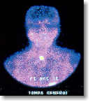 Another Sestamibi scan is shown here on the right. We have used the computer to color enhance the x-ray, thus it is more blue than the other scan. Once again, you can see that this patient has one "HOT" spot in their neck corresponding to a parathyroid tumor that is making too much parathyroid hormone. When performing a MIRP mini parathyroid operation, the little radioactive probe that the surgeon uses in the operating room will find this radioactive tumor quickly. At our center, this tumor would be removed in less than 5 minutes. Another important thing to remember about sestamibi scans is this: Even if it is positive, it does not say anything about the other three parathyroid glands, and at least 15% of people will have a second bad parathyroid gland. This is why we look at all four parathyroid glands in almost every patient because the BEST thing the scan can do is tell what is happening about ONE gland. It says nothing about the other three glands. The video we have online shows this exact situation: the scan shows a beautiful single parathyroid tumor (one tumor only), yet we look at the other 3 glands and found two normal glands and a second tumor. You CANNOT simply take out the hot spot parathyroid tumor. If your surgeon is going to operate on you and take out the hot spot tumor and call it a "mini" parathyroid operation, then know you have a 15% chance that you will need a second operation to remove your second tumor because you will not be cured. You must be careful of the surgeon who is only going to remove the one hot-spot tumor! Watch the video and see it for yourself!
Remember, a Sestamibi scan is a VERY safe procedure. There is NO cross-reactivity for other types of x-ray dye, so parathyroid patients with allergies to x-ray dye can have a Sestamibi scan. Also note that the Sestamibi drug used to show the over-active parathyroid gland is the exact same drug that is used to perform cardiac stress tests--it is very safe! ALSO, the type of radioactivity used is the most mild radioactive agent used in all of medicine. You are in no danger and your family can stay with you--it is not dangerous to them either (or your doctor!).
Also... everybody wants to know how to say "sestamibi". It's: Ses - ta - mee' - bee
 Parathyroid surgery and mini parathyroid surgery uses sestamibi scanning for best cure rates. This picture shows the Sestamibi camera. The "x-ray" pictures above are obtained from this camera. The Sestamibi scan will often display the hyperactive parathyroid gland which is causing hyperparathyroidism in about 80 percent of all patients... BUT, when it does show a single hot gland, it is usually correct. However, it will not tell anything about the other three glands. When combined with the probe in the operating room (the MIRP mini parathyroid operation), and examination of the other glands, the cure rate can be over 99%. It takes approximately two hours for most places to perform a Sestamibi scan. Pictures of the neck and chest are usually taken immediately after the injection and again 1.5 to 2.0 hours later.
Note: When sestamibi scans are performed at the Norman Parathyroid Center, it takes only 15 minutes on average to do a scan. We almost never take more than 20 minutes to do a scan. Sometimes it is done in only 10 minutes. Why? Because we do about 13-14 per day... every day. We do about 20% of all the sestamibi scans in the US every year. We have developed several techniques that nobody else in the world does, thus our scans are very clean, crisp, and accurate. Heck, even our patients can read the x-ray correctly about 90% of the time--that's how easy it is. Remember from above, we try very hard to get good "negative" information. Finding the big tumor is easy for us, it's finding the three other glands that takes talent and experience. After more than 17,000 of these operations we know that showing the parathyroid tumor on the scan is NOT the challenge, the challenge lies in assessing the other three glands. "True Negative" information (knowing 100% that there is nothing under the jaw or in the chest) is absolutely the most important information a scan can tell us. We LOVE negative scans... a really good quality negative scan is the best thing we can get. These patients will have a cure rate of 99.9% at our center with an operation that takes less than 20 minutes. This is what we do all day, every day.
When discussing mini-parathyroid surgery, most endocrinologists and surgeons see things two ways: EASY operations which can be done "minimally invasive" because the scan is positive and the surgeon knows where the tumor is, and HARD because the scan is negative and they don't know where the tumor is located--so they will have to do an exploration to find the tumor. There is a reason why thousands of people travel to Tampa for their parathyroid operation every year--because this view of parathyroid surgery is DUMB. Only inexperienced surgeons need a positive scan for a mini-operation! All parathyroid operations are minimally invasive for us because we do not use the sestamibi scan for any decision making. We do the same, mini-parathyroid operation on every single patient regardless of scan results. If it wasn't easy, we couldn't do 12 or more per day with a cure rate near 100%, with everybody going home in an hour or two. Look folks, we can't say this any clearer--it is not about the scan, it is about the experience of the surgeon. It is not about the golf clubs, it is the guy holding the golf clubs. Stop emphasizing the scan! It's not about the scan. Heck, if you come here--please don't get a scan. We'll do it 15 minutes before your operation.
One of the biggest problems with Sestamibi scanning is the variability in scans from hospital to hospital. Sestamibi scans are not like any other type of x-ray test. It doesn't actually use x-rays, so the pictures are extremely dependent upon the skill of the technician. CAT scans, MRIs, and regular x-rays all are very similar throughout the world. It is easy to do these x-rays because of the technology used. All these x-rays are done the same way everywhere and they are all very excellent quality. Because most hospitals and radiology departments see only a few parathyroid patients per year, they do not get many opportunities to perform a sestamibi scan. IMPORTANT! There is a very high correlation between hospitals that do a lot of Sestamibi scanning (more than 100 per year) and their accuracy. Like other aspects of treating parathyroid disease, the experience of the doctors involved makes all the difference! Click Here for more technical details on how this scan is performed at the Hospital for Endocrine Surgery at the Norman Parathyroid Center. This group has published the highest accuracy rates with Sestamibi scanning and are recognized as world experts at this type of scan. Sadly, some very high-profile hospitals, universities and clinics have some of the worst scans in the US. Here is the simple test to see if you have a good scan.... Simply look at it. If you can pick up your scan, hold it up to the light and see your head, neck, and chest (it MUST be clean and crisp) then you have a good scan. You should see the thyroid gland very clearly (looks like a butterfly). It should not be blurry. If your scan is blurry, then it is junk. PERIOD. If your scan is blurry and it is read as negative, then refuse to pay for it. All patients should demand a copy of their sestamibi scan and look at it. If it looks like a bunch of blurs and blobs, then you have just wasted your time. Do not let your doctors make any decisions on your behalf based upon a sestamibi scan. Our scans are crisp and clear--thus a crisp, clear, in focus negative scan is PERFECT! That's what we want.
Since January of 2003 the Norman Parathyroid Center has been collecting data on sestamibi scans from different hospitals around the US. Patients (and doctors) send their x-rays to us to have them evaluated. Our doctors have reviewed over 60,000 sestamibi scans from hundreds of different hospitals from all 50 states, now averaging about 100 scans per week that they review. He has found that most scans are worthless because the radiology tech that did the scan did not know how to do the scan. We evaluate and score each sestamibi scan and rank it for quality (if you are one of Dr Norman's patients, he will share with you his score of your scans). If you come to Tampa for your operation, you will see your scan and you will be able to see your tumor prior to the operation, and how important the "negative" information is--which lets us know 100% that you do not have a second tumor in your chest, or behind your voice box. The "negative" information (on a clear, crisp scan) is the most important part of the scan.
The Sestamibi Quality Scoring Scale goes from 1 to 10, with a score of 10 meaning that all the right things were done and it is a very high quality scan. If they do everything wrong except your name, they get a score of 1 (meaning you wasted your time and money).
The 10 items scored are: 1) patient name and date correct, 2) correct dye dose, 3) correct camera height, 4) camera angle from front and both sides, 5) camera position (should show just a small part of the liver and heart, not all of it!), 6) camera focus (must not be blurry at all!), 7) correct columnation (filter), 8) No pin-hole views (bad!!), 9) no iodine dual-isotope (very bad scans), 10) correct acquisition time.
Look at this table--and then look at your sestamibi scan--it will make sense to you. Scans that get a score of 3 will show the parathyroid tumor only 20% of the time (said differently... you have a tumor in your neck that's about the size of a grape but the scan can't see it 80% of the time because the scan was done incorrectly). If the sestamibi scan is done a little better and gets a quality score of 5, then there is a 75% chance it will show your tumor. If you have this scan done correctly and the scan is a very high quality (getting a score of 9 or 10) it will show your tumor about 97% of the time. If your scan is blurry and was not done correctly, it will be negative... it won't show your tumor 80% of the time. LOOK AT YOUR SCAN! Is it blurry?
| Score of the Scan | Shows the Tumor | Same Patient Gets Scan Done by Dr Norman |
|---|---|---|
| 3 | 20% | 97% |
| 5 | 75% | 97% |
| 9 | 95% | 97% |
The average score for 40,000++ scans done throughout the US between 1/2003 and 1/2010 is 4.3. As you can see from the chart above, this means that about 52% of scans done in the US are NEGATIVE because of the technician doing the scan--and the protocol that the technician uses. This is BLURRY negative and worthless... this is not crisp, clean negative which has tremendous value. Again, you have a tumor but the x-ray does not show it because they did the scan wrong! Its not always his/her fault, they read the instructions out of a book and the instructions are bad. Thus... if your scan is Negative, it does NOT mean you have 4 bad glands! (extremely important concept!). It means that you most likely had a very poor quality scan. Over 90% of scans that are "negative" at some other hospital are positive when done by Dr Norman's staff. Sound too good to be true? We wish it was not. We wish everybody had good scans. We can't operate on everybody. We can't scan everybody. We are trying to teach, but its hard! You--the patient--need to demand excellence. If your scan is blurry, it is junk and you should demand better! There is nobody out there that makes sure your x-rays are done correctly... its up to you ! Don't pay for a blurry scan.
IMPORTANT: Now that you understand the quality issue of sestamibi scans you will be able to understand the biggest errors that doctors make. If your doctor tells you any of the following...then they are WRONG:
-
"Your scan is negative so you must have 4 bad glands and not just one bad gland." This is rubbish. Run from this doctor! RUN, RUN, RUN! You are about to be treated incorrectly!
-
"Your scan is negative so we should just wait for a while and see what happens." This is rubbish. The scan is negative because some technician doesn't know how to do the scan, or because the parathyroid tumor is attached to the back side of the thyroid (where it is supposed to be) and you can't see it because the thyroid is in the way. Scan results should never be used to decide who gets surgery and who does not. If you have parathyroid disease then you need to get it fixed. Wait to have a stroke? Wait to get severe osteoporosis? High blood pressure? Depression? Chronic fatigue? Memory loss? Wait around so your risk of breast cancer doubles, and the risk for prostate cancer nearly triples? Wait for what? Geeezzzz... because some dude can't do a good x-ray? Watch the video above on this page, it shows a man that had a negative scan so he didn't have surgery... only to develop bad osteoporosis, kidney stones, GERD, and have a heart attack from all the calcium in his coronary arteries.
-
"Your scan is negative so you can't have mini-surgery." This may or may not be true--it depends on the surgeon and his/her expertise with mini-surgery. At our center we perform mini-surgery on ALL patients, regardless of scan results. If you have a surgeon that uses the word "explore", then get out and go somewhere else. If your kids weren't home at midnight on a Friday night you wouldn't start exploring the entire city looking for them. You would know the top 3-4 places where they hang out and you would call them and find them. Watch a video on this topic by Dr Norman--this 8 minute lecture has some great teaching points about finding parathyroid glands during an operation.
If your doctor tells you any of the above, then please print this page and take it to them. Teach them! Remember, our goal here is to spread the word and educate. Parathyroid disease is extremely easy to fix if you have the right personnel. Look folks, it is not about the scan! Scans are way over emphasized by doctors who don't see hyperparathyroidism patients very often. Please, please, stop worrying about the scan... most scans are wrong.
One final thought... some of the worst scans done in the US are done at major universities while some of the best are done at some walk-in radiology clinics. Do not simply believe that your scan is going to be a good scan because you are at some famous big university... Demand to look at the scan. If you can't understand it without anybody explaining it to you... and it looks like a bunch of blobs, or your head is way out of focus and is blurry... then your scan is not going to be helpful. It will confuse everybody..
Demand a copy of your sestamibi scan and look at it yourself. Make sure it is clear and crisp... no blurry scans! No out of focus sestamibi scans! Demand accuracy--its your money, your body, and your health. If you are coming to our Parathyroid Center to have your operation, do NOT get a scan before coming. We will not look at it, and we won't use any information it contains. Don't send your scan to us, we don't want to see it. It is wrong!
What to read next
- Watch video number 1: What is the sestamibi scan, how to read the sestamibi scan, what the scan means.
- Video number 2: Parathyroid glands can't be "anywhere". The importance of "negative" sestamibi information.
- Normal and abnormal parathyroid Function.
- Technical details of How Sestamibi Scans are Performed at the Norman Parathyroid Center (they do more than 2200 per year and developed many of the techniques used for this test).
- View a Movie of a Mini-Parathyroid Operation. Click Here to see the archived movie of this operation as it happened live. There is no blood and the entire operation takes 13 minutes. This is the state of the art in parathyroid treatment.
- How to become our patient