
Preoperative testing to find a parathyroid tumor is now considered 'standard of care'. In other words, virtually all patients should have a localizing study prior to having parathyroid surgery. The Sestamibi scan is discussed in several other areas of this large parathyroid web site--which is undoubtedly the preferred parathyroid test. If this is the first page you have read regarding the tests used to find diseased parathyroid glands, then PLEASE read our "Finding Bad Parathyroid Glands" page FIRST.
HOWEVER, you must understand that these scans are wrong more than they are right. Listen up folks, sestamibi scans are usually wrong, and even when positive are wrong 60% of the time. Be careful with the information that your doctor gets about a scan... it is probably wrong, not right!
Note: If a Minimally Invasive Parathyroid Surgery (which is performed in an outpatient setting) is planned, then the only preoperative test necessary is a Sestamibi scan the morning of the operation. You can bet on this... if you get more than one scan, then your doctor (surgeon) is not an expert.
Do I need this test? The following tests are performed very infrequently and under very specific and unique circumstances.
Use of CAT scan to Find an Overactive Parathyroid Gland
CAT scanning has always been a poor localizing test for enlarged parathyroids. There are currently very rare indications for the use of CAT scans in this setting and this test should never be performed routinely. We have no good pictures to show because they don't exist! Please don't fall for the hype and get a "4-D" CAT scan. And if you do get one and decide to come to our center for your surgery, don't bother sending us the CAT scan. We will not look at it.
Use of MRI scan to Find an Overactive Parathyroid Gland
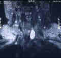 Theoretically, the MRI scan is more sensitive and specific than the CAT scan when looking for enlarged parathyroid glands. Since many endocrine tumors enhance on a T-2 weighted MRI scan this is theoretically preferred over CAT scanning. The photo shows a T-2 weighted MRI of a parathyroid adenoma in the lower left neck within the tracheo-esophageal groove. The indications for MRI scanning for parathyroid glands, however, are still extremely uncommon. It is our strongest opinion that there is almost never an indication for an MRI in patients with hyperparathyroidism. It almost never works and insurance companies should never pay for it. We consult on over 3000 patients with hyperparathyroidism per year and we have NEVER ordered an MRI. This test does not work!
Ultrasound to Find a Bad Parathyroid Gland
Ultrasound is an inexpensive and non-invasive way to look for parathyroid adenomas. Although it is less sensitive and specific than the MRI scan, it is still on occasions because it is readily available, fast and low in cost. Again, most patients do not need any of the localizing studies discussed on this page (CAT scanning, MRI, or ultrasound). Prior to Minimally Invasive Parathyroid Surgery, a Sestamibi scan is mandatory. Since about 1995, the need for using any scanning method other than sestamibi is extremely infrequent. Ultrasound fails to find a parathyroid adenoma half of the time if your endocrinologist or surgeon does the scan himself, but it fails to find the parathyroid tumor 82% of the time if you get the scan done at a radiology clinic. Therefore, it may be cheap, easy, painless, and safe, but its not very good and is usually a waste of time and money (unless done by the endocrinologist). Even when it does "suggest" the location of an adenoma, it is wrong 20 percent of the time, therefore, it is not accurate enough to allow a surgeon to take out this one gland without examining all three other parathyroids. Therefore, ultrasound will not allow a small operation or the use of local anesthesia. Don't fall for the hype. If ultrasound worked great, we'd be doing it every day... but we use it about once per year (one per 3000 cases).
Sestamibi Scanning is the preferred method to find diseased parathyroid glands. We have several pages discussing parathyroid scanning using Sestamibi. However, as noted above, sestamibi is wrong more often than it is right! If a sestamibi scan is negative, it is wrong 100% of the time. When a sestamibi scan is positive, it is wrong 60% of the time. Far too much emphasis is placed on scans. Stop getting scans folks, and stop going to doctors who keep getting scans--it means they don't know what to do. They feel better and more confident if the scan is positive--however, even when it is positive it is wrong more than it is right. Stop putting so much emphasis on the scans folks!